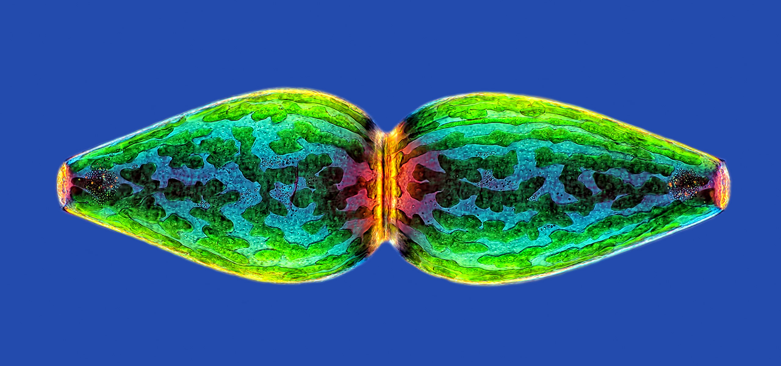
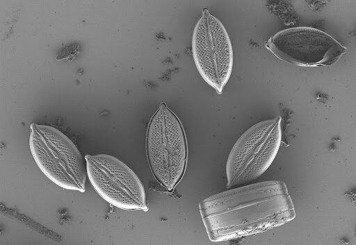
Water through the microscope and the camera
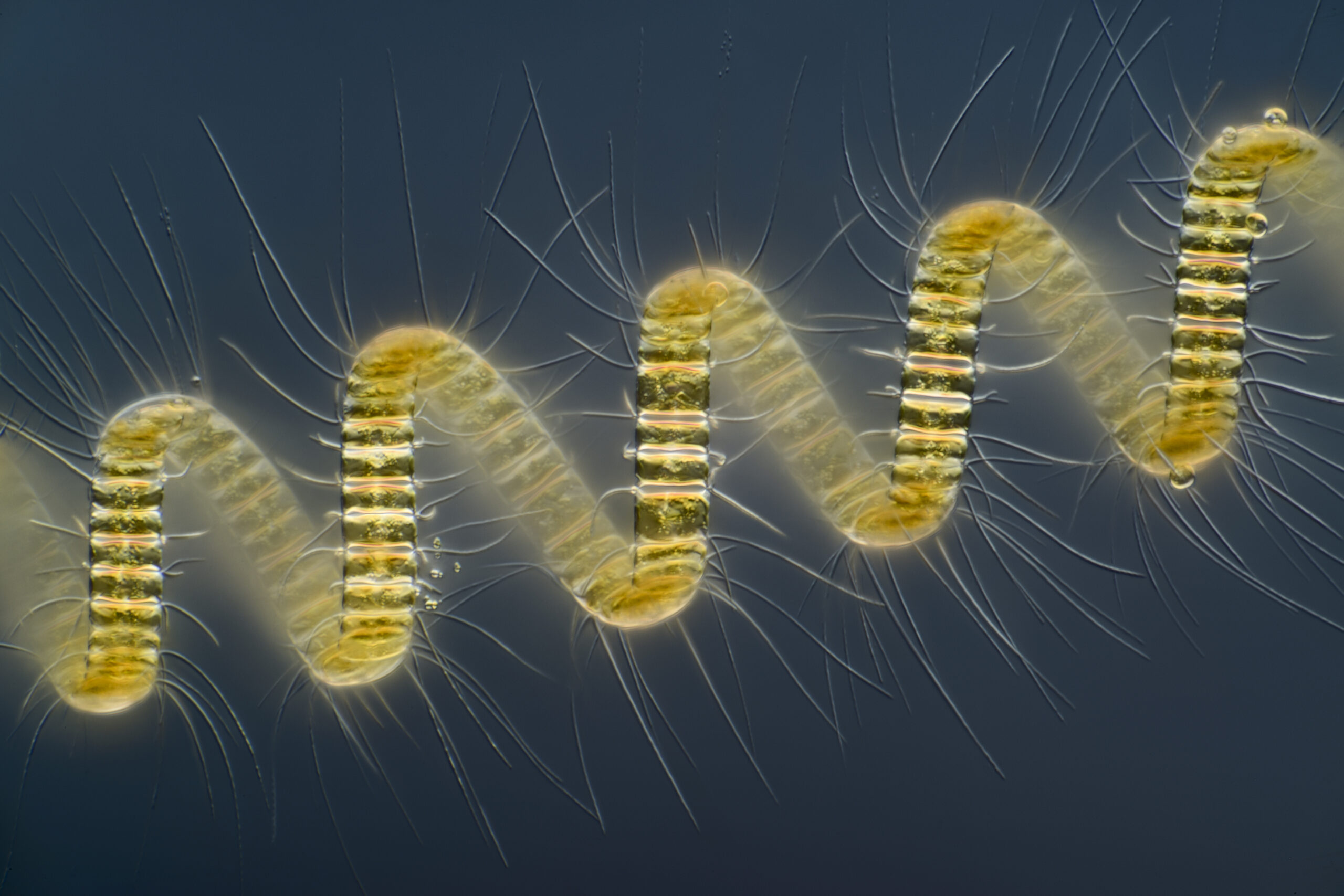
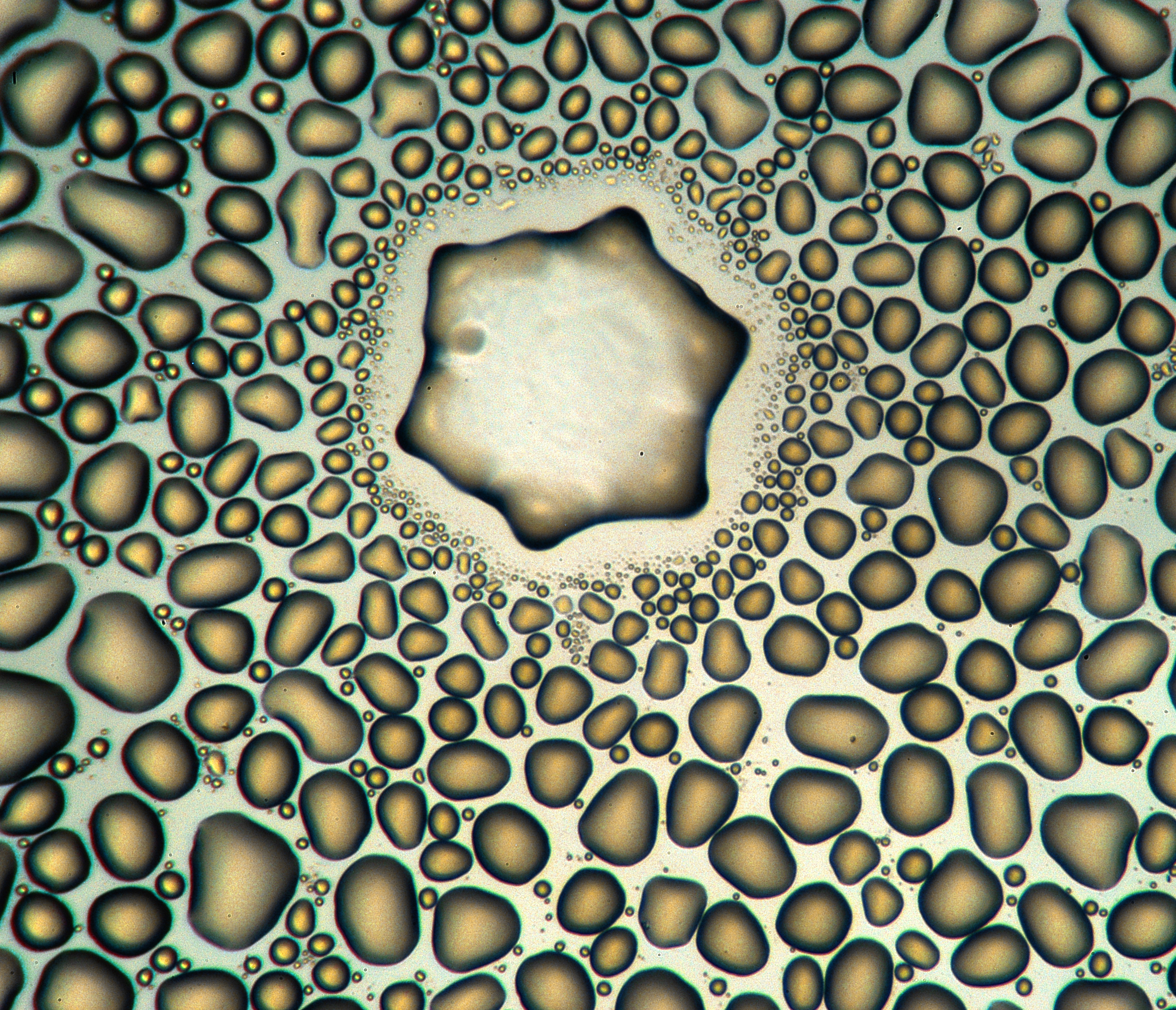
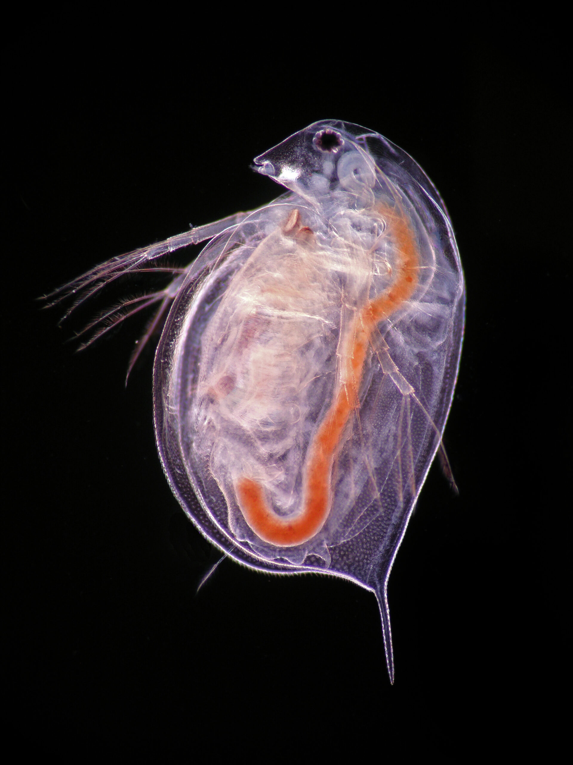
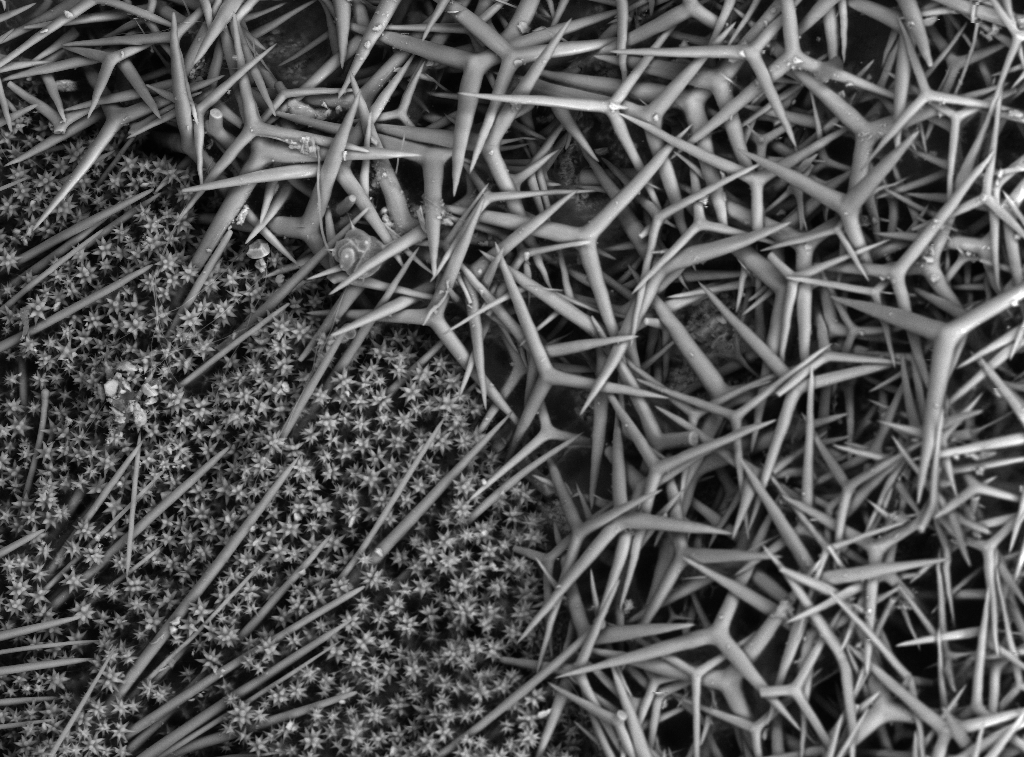
The exposition “Water through the microscope and the camera” was held in March 22, 2017 at the Plenum Gallery, Catania to celebrate the World Water Day 2017. The popular science event was dedicated to water and microscopic life, which is only visible through a microscope and could be captured by the camera. The wall images and video presentation and the scientific seminar revealed the life hidden in a drop of water, combining science, art and photography
MicrobEco promoted knowledge about microorganisms in water to the public of non-experts. Seeing the hidden beauty of Nature, people could better appreciate nature, be less victim of misinformation and elaborate their opinion about environmental and health policies
The Scientific Committee includes personalities from the academics and not: Manuela Coci (MicrobEco) , Gian Marco Luna (CNR-ISMAR-Ancona), Stefano Amalfitano (CNR-IRSA), Angelina Lo Giudice (CNR-IAMC), Antonietta Rosso (Università di Catania), Chiara Pennesi (Università Politecnica delle Marche), Grazia Quero (CNR-ISMAR-Venezia), Fabrizio Joppolo (Nikon Instruments), Michele Nubile (Mima srl), Franco Ferro, Alberto Castro (Plenum Gallery).
Patronage and sponsors: Academia Gioenia of the University of Catania, CNR – ISMAR (Institute for Marine Sciences), CNR IRSA (Institute for Water Research), United Nation- World Water Day, SciencED Nikon and Nikon Small World photomicrography Competition, NPI-Italian S.r.l.- a Neoperl Company, A2D-Analisi Acqua a Domicilio, Plenum Gallery, MIarte exclusive.
PHOTO EXPOSED WITH THE PERMISSION OF NIKON SMALL WORLD